

Introduction
The brain is an organ of immense complexity, requiring a robust blood supply to function correctly. Cerebral circulation is essential for delivering oxygen and nutrients while removing metabolic waste. This article explores the anatomy of the brain’s arterial supply, touches on venous drainage, and discusses important clinical aspects, including intracranial aneurysms, cerebral vasospasm, and the implications of the Circle of Willis in anesthesia.
The Arterial Supply
The brain’s blood supply is provided by four major arteries: two internal carotid arteries and two vertebral arteries.
Internal Carotid Arteries
The internal carotid arteries provide around two-thirds of the brain’s blood supply. These arteries give rise to the middle cerebral arteries, supplying the lateral parts of the cerebral hemispheres and much of the internal capsule. The internal carotids also give rise to the anterior cerebral arteries, which are connected by the anterior communicating artery and supply the medial and superior aspects of the hemispheres.
Vertebral Arteries and the Basilar Artery
The vertebral arteries contribute the remaining one-third of the brain’s blood supply. Each vertebral artery gives off the posterior inferior cerebellar arteries before joining to form the basilar artery. The basilar artery gives rise to the anterior inferior cerebellar and the superior cerebellar arteries.
The basilar artery then splits into the two posterior cerebral arteries, which supply the medial side of the temporal lobe and the occipital lobe. These posterior cerebral arteries connect with the carotid arteries via the posterior communicating arteries.
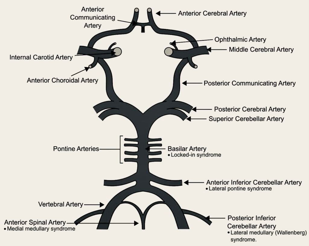
The Circle of Willis
These major arteries are interconnected by a structure known as the Circle of Willis, formed by the anterior and posterior communicating arteries. This arterial circle provides a vital collateral blood flow pathway, although it’s worth noting that in up to 15% of normal asymptomatic individuals, the Circle of Willis is incomplete.
The Venous System
Venous drainage of the brain, though less often highlighted, is equally important.
Dural Sinuses
The cerebral and cerebellar cortices drain into the dural sinuses, which lie between the two layers of the cranial dura mater. The superior sagittal sinus, running along the attached edge of the falx cerebri, typically drains into the right transverse sinus. The inferior sagittal sinus, found along the free edge of the falx, drains into the left transverse sinus via the straight sinus. The transverse sinuses merge into the sigmoid sinuses, which exit the cranium as the internal jugular veins.
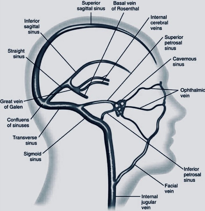
Deeper Structures
Deeper structures of the brain drain through the internal cerebral veins, which combine to form the great cerebral vein, or the vein of Galen. This vein also drains into the inferior sagittal sinus. The cavernous sinuses, situated on either side of the pituitary fossa, eventually drain into the transverse sinuses.
Implications of the Circle of Willis in Anesthesia
The Circle of Willis, an essential structure in the brain’s arterial supply, plays a significant role in anesthesia.
- Collateral Circulation: The Circle of Willis provides alternative pathways for blood flow in case of an arterial occlusion. During surgeries that involve manipulating major arteries, such as carotid endarterectomy or intracranial procedures, a robust Circle of Willis ensures that the brain can still receive adequate blood supply even if one or more vessels are temporarily obstructed.
- Ischemia Prevention: During anesthesia, particularly in scenarios where induced hypotension is used to reduce surgical bleeding, the Circle of Willis helps maintain cerebral perfusion. Its ability to distribute blood flow from patent arteries to areas at risk of reduced perfusion helps mitigate the risk of brain ischemia.
- Surgical Planning: An understanding of the Circle of Willis is integral to planning neurovascular surgeries. Surgeons can devise strategies that minimize the risk of ischemic events by ensuring that blood flow through collateral pathways is maintained. Preoperative imaging to assess the completeness and functionality of the Circle of Willis can inform decisions on surgical approaches, timing, and techniques.
- Aneurysm Management: The Circle of Willis is a common site for intracranial aneurysms. Knowledge of its anatomy is essential for diagnosing and planning the treatment of these aneurysms. Endovascular coiling or surgical clipping requires precise navigation of this complex vascular network.
- Monitoring Cerebral Perfusion: During anesthesia, continuous monitoring of cerebral perfusion pressure is vital to ensure adequate oxygenation of the brain. The Circle of Willis facilitates this by distributing blood flow from multiple arterial sources. Anesthesiologists can use transcranial Doppler ultrasonography and other monitoring techniques to assess the integrity and functionality of the Circle of Willis.
- Understanding Variations: Anatomical variations in the Circle of Willis are common and can influence anesthetic management. Incomplete circles or variations in vessel size and connectivity can alter the distribution of blood flow. Preoperative imaging studies, such as MR angiography, can reveal these variations and guide perioperative management strategies.
- Endovascular Procedures: The Circle of Willis is a critical landmark for endovascular interventions. During procedures like angioplasty, stenting, or thrombectomy, accurate placement of catheters and devices depends on a clear understanding of this vascular network.
- Hemodynamic Stability: Maintaining hemodynamic stability is paramount during anesthesia, especially in patients with cerebrovascular disease. The Circle of Willis’s role in providing collateral blood flow underscores the importance of avoiding significant fluctuations in blood pressure.
- Anesthetic Techniques: Anesthetic techniques must be carefully chosen to minimize their impact on cerebral blood flow. Agents that preserve autoregulation and avoid significant vasodilation or vasoconstriction are preferred.
- Patient Positioning: Patient positioning during surgery can influence cerebral blood flow dynamics. Understanding the anatomy of the Circle of Willis helps anesthesiologists and surgeons position patients in ways that optimize perfusion.
Conclusion
The cerebral circulation, encompassing both the arterial supply and venous drainage, is a complex and vital system. Understanding its anatomy and clinical implications, particularly the role of the Circle of Willis, is crucial for managing various medical and surgical scenarios. This knowledge helps ensure adequate cerebral perfusion, prevent ischemic events, and improve patient outcomes during anesthesia and surgery.