
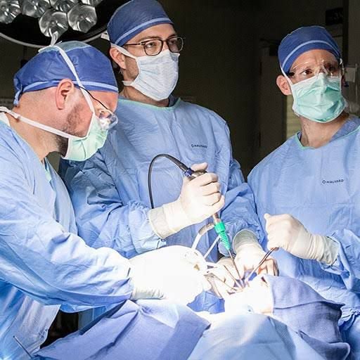
The field of neuroanesthesia has witnessed significant advancements in the past two decades, aligning itself with the rapid technological developments in neurosurgery. These advancements have paved the way for novel surgical approaches and procedures, which, in turn, have introduced new challenges in the perioperative care of neurosurgical patients. Notably, the aging population, characterized by an increase in life expectancy, has led to a growing number of geriatric patients seeking neurosurgical interventions. However, the functional impact of surgery, coupled with the presence of comorbidities, can pose additional complexities, potentially affecting patient outcomes.
This article delves into the evolving landscape of neuroanesthesia, highlighting the interplay of technology, surgical techniques, and patient demographics. We will explore the role of minimally invasive surgical practices that have flourished in tandem with advancements in computer-assisted technology, imaging modalities like computed tomography (CT) and magnetic resonance imaging (MRI), and innovative surgical instruments. These developments have enabled surgeons to achieve a three-dimensional (3-D) visualization of the surgical site, enhancing precision while minimizing tissue damage.
Moreover, stereotactic systems, integrating patient co-registration with preoperative imaging data, have revolutionized the field, enabling surgeons to navigate instruments with pinpoint accuracy. For geriatric patients, these technological innovations translate into less invasive procedures, reduced tissue trauma, enhanced precision, decreased pain, expedited recovery, and improved overall outcomes.
Fiberoptics have played a pivotal role in revolutionizing visualization within the surgical field. The utilization of specialized endoscopes, inserted through small incisions, permits visualization of hidden areas with minimal dissection, reduced pain, shorter surgical durations, minimal scarring, and quicker recovery times. While endoscopic procedures offer numerous advantages, they do present a limitation: depth perception. However, ongoing advancements, such as newer and more costly 3-D endoscopy, hold the potential to overcome this limitation.
Furthermore, the integration of robotic technology into neurosurgery has facilitated minimally invasive procedures. Robotic-assisted neurosurgery involves the use of robotic arms to enhance surgical precision and access, leading to smaller incisions, reduced blood loss, shorter operating times, earlier recovery, and shorter hospital stays.
Endoscopic Transnasal Transsphenoidal Hypophysectomy
Advantages of Endoscopic Approach for Pituitary Tumor Resection
| Advantages | Description |
|---|---|
| Broader and clearer field visualization | Enhanced visibility of the surgical area |
| Ability to access hidden areas | Improved access to regions not visible with a microscope |
| Less pain | Reduced postoperative discomfort |
| Shorter operative times | Faster surgical procedures |
| Better airflow | Improved breathing during and after surgery |
| Earlier recovery | Quicker recuperation post-surgery |
| Shorter hospital stay | Reduced duration of hospitalization |
| Better short-term sinonasal quality of life | Enhanced quality of life in the short term |
| Improved endocrinological outcome | Enhanced hormonal function post-surgery |
Anesthesia Considerations
| Consideration | Description |
|---|---|
| Patient Immobility | Ensure complete patient immobility during surgery. |
| Intracranial Pressure (ICP) Monitoring | Vigilance for sudden increases in intracranial pressure. |
| Prompt Neurological Examination | Plan for early emergence from anesthesia to conduct a neurological exam. |
| Geriatric Patients | Evaluate for a history of hypertension, diabetes, CAD, compromised renal function, etc. |
| Drug History | Assess the patient’s drug history. |
| Evaluation of Organ Systems | Properly evaluate all organ systems. |
| Difficulty in Mask Ventilation | Loss of dentition may make mask ventilation difficult. |
| Difficulty during Laryngoscopy | Arthritic changes in the spine may cause difficulties during laryngoscopy. |
| Nasal Cavity Decongestion | Use cotton pads soaked in an adrenaline and local anesthetic mixture for decongestion. |
| Blood Pressure Control | Strictly control blood pressure to avoid bleeding that may obscure the operating field. |
| Anesthesia Aids for Blood Pressure Control | Use Propofol or dexmedetomidine infusion to regulate blood pressure and prevent sudden increases. |
| Nerve Blocks | Consider maxillary, infraorbital, sphenopalatine, and pterygopalatine nerve blocks as adjuvants to general anesthesia for hemodynamic control and analgesia. |
Comparison of Postoperative Complications: Endoscopic vs. Microscopic Techniques
| Complication | Incidence in Endoscopic Techniques | Incidence in Microscopic Procedures |
|---|---|---|
| Diabetes Insipidus | Higher | Lower |
| Syndrome of Inappropriate ADH Secretion | Higher | Lower |
| Cerebrospinal Fluid (CSF) Rhinorrhea | Higher | Lower |
| CSF Leak | Higher | Lower |
| Fever | Higher | Lower |
Please note that the comparison of postoperative complications between endoscopic and microscopic techniques may vary in different studies.
Endoscopic Third Ventriculostomy
Procedure Description
Endoscopic third ventriculostomy is a minimally invasive surgical technique used to treat conditions that result in the blockage of cerebrospinal fluid (CSF) flow within the brain’s ventricular system. During this procedure, an endoscope is inserted through a small burr hole into the ventricular system and directed toward the third ventricle. In the third ventricle, a surgical opening is created using the endoscope. This opening allows CSF to flow directly into the basal cisterns, bypassing any obstructions beyond the third ventricle.
Anesthetic Management
| Anesthetic Management Consideration | Description |
|---|---|
| Preoperative Optimization | Similar to patients with elevated intracranial pressure, preoperative management focuses on correcting fluid and electrolyte abnormalities. Sedatives are usually avoided in premedication. |
| Anesthetic Induction | Standard anesthetic induction is followed by endotracheal intubation. Anesthesia can be maintained with air/inhalational agents or total intravenous anesthesia. |
| Nitrous Oxide Avoidance | Nitrous oxide is avoided due to concerns about the expansion of air bubbles in the ventricular system. |
| Monitoring | Standard American Society of Anesthesiologists (ASA) monitoring includes ECG, blood pressure, pulse oximetry, capnography, and urine output monitoring. Arterial line monitoring allows beat-to-beat blood pressure monitoring for hemodynamic management. |
| Intraoperative Arrhythmias | Intraoperative rate arrhythmias may occur due to elevated ICP or stimulation of adjacent structures like the hypothalamus and brainstem. |
| Basilar Artery Awareness and Injury | The basilar artery, located beneath the floor of the third ventricle, may rarely be injured, leading to massive hemorrhage. |
| Intraoperative Irrigation | Continuous fluid irrigation is performed during neuroendoscopy of ventricles. This can result in increased ICP and decreased cerebral perfusion pressure (CPP). Monitoring pressure inside the neuroendoscope helps manage ICP. |
| Irrigation Fluid Choice | Warm Ringer lactate is commonly used as an irrigation fluid to avoid temperature dysregulation and hypothermia. |
| Perioperative Complications | Possible complications include delayed awakening, pneumocephalus, infection, and convulsions. Blood transfusion is generally not required intraoperatively unless massive hemorrhage occurs, which is rare. |
| Postoperative Extubation | The trachea is extubated postoperatively in uncomplicated cases. Delayed awakening may occur in some cases. |
It’s important to note that endoscopic third ventriculostomy is a specialized procedure performed to treat specific conditions, and the choice of anesthesia and management should be tailored to individual patient needs and surgical requirements.
Endoscopic Ventriculoperitoneal Shunts
Procedure Description
Ventriculoperitoneal (VP) shunt placement is a surgical procedure used to treat conditions involving the abnormal accumulation of cerebrospinal fluid (CSF) within the brain’s ventricular system. In this procedure, a shunt system is inserted to divert excess CSF from the ventricles to the peritoneal cavity, where it can be reabsorbed.
Anesthetic Management
| Anesthetic Management Consideration | Description |
|---|---|
| Intraoperative Guidance | Intraoperative use of ultrasound or frameless stereotaxy for placing the ventricular end of the shunt improves catheter placement accuracy and reduces proximal shunt failures. |
| Laparoscopic Approach | Minimally invasive laparoscopic insertion of the distal catheter is compared to conventional laparotomy. It reduces operating times, lowers shunt failure rates, and minimizes the risk of abdominal malposition. |
| Anesthetic Implications | There are no significant differences in anesthetic implications between the minimally invasive laparoscopic approach and the conventional freehand technique for VP shunt placement. |
The choice of the technique used for VP shunt placement may depend on the specific clinical scenario and surgeon preference. However, the anesthetic management principles remain consistent, focusing on ensuring patient safety and optimizing outcomes.
Aneurysm Clipping: Minimally Invasive Approaches
Aneurysm Clipping Overview
Aneurysm clipping is a surgical procedure used to treat cerebral aneurysms, which are abnormal, balloon-like bulges in the walls of brain arteries. Traditional pterional craniotomy, while effective, has associated complications and disadvantages, leading to the development of minimally invasive approaches.
| Surgical Approach | Description |
|---|---|
| Pterional Craniotomy | The classic pterional craniotomy involves significant retraction of the temporalis muscle. It can lead to complications such as delayed atrophy/scarring, facial asymmetry, temporomandibular joint dysfunction, pain on mastication, and injury to the frontal branch of the facial nerve. This approach exposes a large area of cerebral cortex. |
| Minimally Invasive Microsurgery | Minimally invasive microsurgery techniques, including keyhole or mini-craniotomies, have gained popularity due to shorter operative times, reduced pain, improved cosmesis, lower cost, and enhanced patient acceptability. These approaches minimize the drawbacks associated with the classic pterional craniotomy. |
| Endoscope-Assisted Surgery | Endoscope-assisted aneurysm surgery utilizes an endoscope for various purposes: (a) inspection before clipping to visualize hidden areas, (b) clipping under endoscopic view, or (c) post-clipping evaluation to ensure complete clipping and perforator preservation. Purely endoscopic aneurysm surgery is not widely adopted. |
The shift toward minimally invasive microsurgery and endoscope-assisted techniques in aneurysm clipping is driven by the desire to reduce complications, enhance patient outcomes, and improve overall surgical experience. However, the choice of approach depends on individual patient factors and surgeon expertise.
Stereotactic Procedures in Neurosurgery: Anesthetic Considerations
Stereotactic Procedures Overview
Stereotactic procedures play a crucial role in neurosurgery by providing precise guidance for minimally traumatic interventions. There are two main approaches to stereotactic procedures: frame-based and frameless stereotaxy.
| Stereotactic Approach | Description |
|---|---|
| Frame-Based Stereotaxy | Utilizes a fixed rigid metallic ring frame with fixed fiducials and an instrument holder. Involves brain imaging and computerized superimposition to obtain a 3-D image for guidance. Often used for procedures like deep brain stimulation (DBS). |
| Frameless Stereotaxy | Avoids the use of a rigid frame, instead relying on scalp markers as fiducial points. These markers help relate the computer-generated 3-D image to surgical instruments. Commonly used for procedures such as brain tumor biopsy or excision, abscess/hematoma drainage, and DBS. |
Anesthetic Management for Deep Brain Stimulation (DBS)
DBS is a critical procedure typically performed on elderly patients with Parkinson’s disease. Anesthetic goals focus on providing optimal surgical conditions, patient comfort, managing intraoperative complications, and facilitating intraoperative neuromonitoring.
| Key Anesthetic Considerations for DBS |
|---|
| Elective Awake Surgery |
| – The initial stage involves electrode placement at the target nuclei of the basal ganglia. Patients are kept awake and cooperative during this phase for microelectrode recordings (MER). Anesthetics that affect GABA action are generally avoided. Dexmedetomidine can be considered for sedation. |
| Airway Preparedness |
| – Multiple options for securing the airway should be readily available in the operating room, including a wrench to open the frame if necessary. |
| Patient Positioning |
| – Patients should be positioned comfortably with soft padding. Invasive blood pressure monitoring and measurement of urinary output are carried out. |
| Proper Sedation |
| – Sedatives are discontinued to ensure patients are awake and cooperative during brain stimulation. Alterations in motor symptoms during stimulation help with correct stimulator lead placement. |
| General Anesthesia (if required) |
| – For uncooperative patients or those with severe movement disorders, general anesthesia (GA) is administered. Electrode placement is guided by MRI under GA. |
| Intraoperative Complications |
| – Potential complications include airway obstruction, respiratory dysfunction, hypertension, orthostatic hypotension, venous air embolism, seizures, and loss of cooperation. |
| Second Stage – Battery Placement |
| – This stage is performed under GA using a laryngeal mask airway or endotracheal tube. Postoperative concerns include nausea, vomiting, exacerbation of off-drug state, and rare complications like intracerebral hemorrhage or pneumocephalus. |
Stereotactic Radiosurgery (SRS)
SRS is a noninvasive technique that focuses radiation on a specific pathological area to destroy it. Patients are usually awake but may receive general anesthesia if they are uncooperative or have movement disorders.
SRS is commonly used for conditions such as arteriovenous malformations, acoustic neuromas, and trigeminal neuralgia.
The choice between awake and general anesthesia for SRS depends on patient cooperation and tolerance to immobility during the procedure.
Minimally Invasive Spinal Procedures for Geriatric Patients
Geriatric patients with multiple comorbidities often require spinal surgeries for various conditions such as prolapsed disc, canal stenosis, spondylolisthesis, spinal tumors, or spinal trauma necessitating instrumentation. In such cases, minimally invasive spinal procedures have gained popularity due to their potential benefits for high-risk patients.
Benefits of Minimally Invasive Spinal Procedures
Minimally invasive spinal procedures, including endoscopy, offer several advantages over conventional open surgery:
| Advantages | Description |
|---|---|
| Reduced Muscle Dissection | Minimal muscle dissection results in less trauma to the surrounding tissues. |
| Preservation of Blood Supply | Decreased injury to epidural blood supply contributes to reduced blood loss during surgery. |
| Shorter Operative Time | Minimally invasive techniques often lead to shorter operative times, reducing anesthesia duration. |
| Decreased Postoperative Pain | Patients experience less postoperative pain, improving overall comfort and satisfaction. |
| Early Mobilization | Faster recovery allows for early mobilization and quicker return to daily activities. |
| Shorter Hospital Stay | Reduced complications and quicker recovery contribute to shorter hospital stays. |
| Improved Cosmesis | Minimally invasive approaches result in better cosmetic outcomes with smaller incisions. |
| Early Functional Recovery | Patients regain functionality sooner, which is especially crucial for geriatric individuals. |
However, it’s essential to consider that minimally invasive spine surgery comes with challenges:
- Challenges of Minimally Invasive Spine Surgery *
- Steep Learning Curve: Surgeons need specialized training and expertise to perform these procedures effectively.
- Patient Selection: Careful patient selection is vital to ensure that individuals are suitable candidates for minimally invasive techniques.
- Anatomical Constraints: Reduced anatomical and visual exposure of nerve roots increases the risk of injury, necessitating intraoperative neurophysiological monitoring.
- Postoperative Delirium: Spine surgery in elderly patients is associated with postoperative delirium, with factors like age, blood loss, operative time, and cerebrovascular disease influencing its occurrence. Minimally invasive techniques may help reduce blood loss and operative time, potentially benefiting in this regard. However, more research is needed on the impact of minimally invasive spinal surgery on postoperative delirium.
Minimally Invasive Surgery for Intervertebral Disc Herniation
Intervertebral disc herniation is a common spinal condition that can be treated through minimally invasive surgical techniques. Three primary approaches for this condition are micro-discectomy using a tubular retractor system, percutaneous laser disc decompression, or selective endoscopic discectomy. These procedures can be performed at various spinal levels, including lumbar, cervical, and thoracic regions.
Minimally Invasive Techniques for Intervertebral Disc Herniation
- Micro-Discectomy Using Tubular Retractor System: This technique involves the use of a tubular retractor system to access the herniated disc. It offers a minimally invasive way to remove the herniated portion of the disc.
- Percutaneous Laser Disc Decompression: In percutaneous laser disc decompression, laser energy is used to reduce the size of the herniated disc, alleviating pressure on surrounding structures and nerves.
- Selective Endoscopic Discectomy: Initially used for lumbar discectomy, this technique has expanded to include cervical and thoracic levels. It involves the use of an endoscope to visualize and remove the herniated disc material.
Anesthetic Considerations
The choice of anesthesia for these procedures varies but typically includes:
- General Anesthesia (GA): GA is commonly used and provides complete immobility. However, it may be associated with an increased risk of neurological complications, necessitating intraoperative neurophysiological monitoring.
- Local Anesthesia with Conscious Sedation: For certain cases, local anesthesia combined with conscious sedation is an option. Patients under local anesthesia can communicate with the surgeon, alerting them to any nerve root impingement.
- Spinal/Epidural Anesthesia: In percutaneous endoscopic discectomy, spinal or epidural anesthesia may be chosen, along with local anesthesia and conscious sedation.
- Dexmedetomidine: Intravenous sedative agents like dexmedetomidine have gained favor due to their safer respiratory profiles. However, it’s essential to monitor for respiratory depression, and conversion to GA may be required if needed.
In summary, minimally invasive surgical techniques for intervertebral disc herniation offer various options for anesthesia. The choice depends on patient factors, surgical requirements, and the surgeon’s preferences. Each technique has its advantages and considerations, making patient-specific selection crucial for optimal outcomes.
Endoscopic Microforaminotomy
Endoscopic microforaminotomy is a minimally invasive surgical procedure aimed at relieving pressure on the spinal cord or nerve roots. In comparison to open cervical foraminotomy, this technique offers several advantages, including lower blood loss, reduced pain, and shorter hospital stays. These procedures are typically conducted under general endotracheal anesthesia.
Types of Endoscopic Microforaminotomy:
- Anterior Cervical Foraminotomy: This approach is used to treat ventral radiculopathy without the need for discectomy, fusion, or neck collar. It is especially effective for cases of foraminal stenosis due to osteophytes or lateral disc herniation.
- Posterior Cervical Microforaminotomy: This procedure addresses foraminal stenosis caused by degenerative changes. It can be performed with the patient in either the prone or sitting position.
Anesthetic Considerations:
General endotracheal anesthesia is typically employed for these procedures. Important considerations include:
- Intraoperative evoked potential monitoring, which necessitates total intravenous-based anesthesia without muscle relaxants.
- Potential risks include nerve root injury, vascular damage, or injury to the sympathetic chain, leading to Horner syndrome.
- Patients are typically awakened and extubated at the conclusion of the surgery.
Video-Assisted Thoracoscopic Spine Surgery (VATS)
Video-assisted thoracoscopic spine surgery (VATS) is a growing approach for correcting scoliosis/kyphosis and treating spine trauma in the anterior thoracic and thoracolumbar junction regions. It offers several benefits, including reduced chest wall morbidity, less pain, and improved cosmesis.
Anesthetic Considerations:
- Preoperative evaluation, especially for cardiac and pulmonary conditions, is essential.
- Single-lung ventilation is crucial for optimal visualization during VATS. This is achieved using a double-lumen endotracheal tube.
- Patients are positioned in the lateral decubitus position, and careful attention is given to cervical spine involvement, which is common in elderly patients.
- Nitrous oxide is typically avoided. Intraoperative monitoring includes somatosensory-evoked potentials (SSEPs) and motor-evoked potentials (MEPs).
- Multilevel spine surgery may involve significant blood loss, and any large vessel injury may necessitate conversion to open thoracotomy.
Percutaneous Kyphoplasty and Vertebroplasty
Percutaneous kyphoplasty and vertebroplasty are procedures used to treat vertebral fractures, commonly seen in elderly patients with multiple medical conditions.
- Positioning is crucial due to osteoporosis, and procedures can be done under general anesthesia or local anesthesia with sedation.
- Intravenous midazolam and fentanyl are typically used for vertebroplasty.
- Kyphoplasty may require additional opioid boluses during balloon tamp insertion.
- Dexmedetomidine has been used as an alternative sedative agent.
- Intraoperative extravasation of bone cement to veins or epidural spaces may occur and should be monitored closely.
Minimally Invasive Lumbar Spine Fusion
Lumbar spine stabilization is often necessary for elderly patients with various spinal conditions. Minimally invasive techniques offer reduced muscle and tissue damage, shorter operative times, less blood loss, and faster recovery.
- Procedures include anterior lumbar interbody fusion (ALIF), posterior lumbar interbody fusion, transforaminal lumbar interbody fusion, intertransverse fusion, and pedicle screw/rod placement.
- ALIF can be done laparoscopically through transperitoneal or retroperitoneal routes. Retroperitoneal fusion should consider laparoscopic surgery-related concerns.
- Transforaminal, posterolateral procedures, and percutaneous spinal fixation are conducted in the prone position.
Anesthesia selection and perioperative management depend on patient-specific factors, surgical requirements, and the surgeon’s preferences for each of these minimally invasive spine procedures.