
- Stereotactic Electroencephalography (SEEG) is an invasive method used to monitor and localize seizure foci in drug-resistant focal epilepsies.
- It involves the implantation of leads to record seizures and achieve three-dimensional analysis of the epileptogenic zone.
- The history of SEEG dates back to the 19th century, with advancements in understanding the electrical activity of nerves, muscles, and the brain.
- Intra-operative EEG was first recorded by Penfield and Jasper, who also used electrocorticography (ECoG) for epilepsy treatment.
- SEEG was introduced in the 1950s by Talairach and Bancaud, using stereotactic techniques for precise electrode placement.
- The use of stereotactic devices, angiography, and later MRI improved accuracy in locating targets for electrode placement.
- Anesthesia for SEEG has evolved to ensure it doesn’t interfere with intraoperative EEG monitoring, with anesthesiologists selecting appropriate drugs for induction and maintenance.
- Various forms of intraoperative EEG monitoring have been attempted, but none has been proven superior to others.
Indication of Stereo-Electroencephalography (SEEG):
- SEEG is indicated for patients with medically refractory focal epilepsies who are candidates for surgical treatment.
- Collaboration with noninvasive preoperative investigations like imaging and video-EEG may not always provide conclusive results, necessitating the use of invasive techniques like SEEG.
- Specific criteria for choosing SEEG over other invasive methods include deep-seated or challenging-to-reach epileptogenic zones, previous subdural study failure, extensive bi-hemispheric exploration, and indications of functional network involvement despite normal MRI findings.
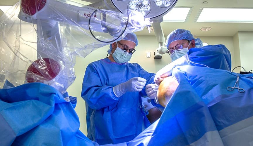
Surgical Techniques of SEEG Electrode Implantation:
- Preoperative MRI with stereotactic neuro-navigation software is used to calculate trajectories.
- Leksell stereotactic frame is applied to the patient under general anesthesia.
- Stereotactic Dyna CT and 3D digital subtraction angiography may be performed to ensure accuracy.
- Fused images from preoperative MR, Dyna CT, and angiographic scans guide electrode placement.
- Depth electrodes are used to reach the desired targets with live fluoroscopic control.
- Post-implantation Dyna CT scans are obtained to confirm electrode placement accuracy.
Robotic SEEG Placement:
- This technique involves precise positioning of electrodes using a surgical robot, like the Renishaw neuromate®.
- Gadolinium-enhanced MRI is used to locate surface vessels.
- Trajectories are planned using robot software.
- The Leksell stereotactic frame is used for preoperative localization.
- Robotic arm-driven electrode placement is monitored, and immediate postoperative CT scans are acquired for validation.
Pre-operative Assessment:
- Patients with intractable epilepsy are often on long-term, multiple anti-epileptic medications, which can affect anesthetic management.
- Anti-epileptic drug interactions, potential side effects, and modifications should be considered.
- Anti-platelet medications should be discontinued before surgery.
- Patients on long-term anticoagulation therapy require careful peri-operative planning.
Peri-operative Monitoring:
- Standard monitoring per guidelines of organizations like ASA and AAGBI should be established.
- Invasive arterial blood pressure monitoring may be necessary in certain cases.
- Neuromuscular blockade can be monitored using a peripheral nerve stimulator to evaluate the Train of Four ratio.
Anesthetic Goals and Peri-Operative Anesthetic Management:
- Anesthetic goals for SEEG include smooth induction and emergence, maintaining adequate cerebral perfusion pressure, ensuring absolute patient immobility, minimizing interference with intra-operative EEG monitoring, enhancing the chance of seizure detection, and addressing complications.
Effect of Different Anesthetic Medications on EEG:
- Anesthetic drugs like Propofol, thiopental, Isoflurane, and sevoflurane can impact EEG patterns.
- Intravenous anesthetic agents like Propofol and Etomidate have dose-dependent effects on EEG.
- Benzodiazepines produce specific EEG patterns at different doses.
- Barbiturates can enhance epileptic activity at low doses.
- Ketamine produces unique EEG changes and may provoke seizures in patients with epilepsy.
- Inhalational agents can affect EEG, with varying degrees of depression depending on the agent.
- Opioids cause dose-related changes in EEG.
- Muscle relaxants generally do not affect EEG.
Special Considerations During Maintenance and Emergence from Anesthesia:
- Maintenance of anesthesia for SEEG often involves a balance between intravenous and inhalational agents.
- Techniques to enhance the chance of seizure detection include using remifentanil infusion, maintaining low concentrations of inhalational agents, and, in some cases, using low-dose Ketamine infusion.
- Smooth emergence is crucial to avoid sympathetic responses and potential complications.
- Careful control of opioids, muscle relaxants, and anesthetic agents is necessary to ensure optimal EEG recording conditions.
COMPLICATIONS:
- Inadequate Sedation or Anesthesia: One of the primary responsibilities of an anesthesiologist during SEEG is to provide optimal sedation or anesthesia. Inadequate sedation can lead to patient discomfort and movement, which may interfere with electrode placement. On the other hand, excessive sedation can complicate the neurophysiological monitoring. Balancing the depth of anesthesia is crucial.
- Infection: Like any surgical procedure, SEEG carries a risk of infection. Anesthesiologists must ensure strict adherence to aseptic techniques and the use of appropriate antibiotics to minimize this risk. Any signs of infection, such as fever or worsening pain, should be promptly investigated.
- Hemorrhage: Intracranial hemorrhage is a rare but serious complication. Anesthesiologists should monitor blood pressure closely to prevent hypertension, which can increase the risk of bleeding. Additionally, careful coordination with the neurosurgeon is essential during electrode placement to avoid vascular structures that may lead to hemorrhage.
- Seizures: SEEG involves inducing seizures for mapping purposes. However, in some cases, seizures can become prolonged or generalize, leading to status epilepticus. Anesthesiologists should be prepared to manage seizures promptly, typically with benzodiazepines or propofol, until seizure termination.
- Neurological Deficits: In rare instances, SEEG electrodes can cause neurological deficits, such as weakness or sensory changes. Anesthesiologists should be vigilant in monitoring for any immediate postoperative neurological changes and communicate them to the surgical team for timely intervention.
- Vessel Injury: During electrode placement, there is a risk of damaging blood vessels. Excessive bleeding can compromise brain perfusion, necessitating rapid hemostasis and blood pressure management by the anesthesiologist.
- Intraoperative Imaging Complications: In some cases, intraoperative imaging, such as CT scans, is used to confirm electrode placement. Anesthesia providers should be prepared for these procedures, ensuring the patient’s safety during transportation and imaging while maintaining anesthesia.
- Postoperative Pain and Nausea: After SEEG, patients may experience postoperative pain or nausea. Anesthesiologists play a role in providing postoperative pain management and antiemetic medications to enhance patient comfort.